Foot X Ray Pittsburgh

Overview of Extremity Foot X Ray Imaging
By definition, an extremity foot X Ray is a diagnostic image of the bones of the body’s extremities. The joints and small bones found here are some of the most complex areas of the body. As such, considerable skill is needed to take and interpret these Foot X Ray images. Extremities include the hands, wrists, hips, knees, ankles, and feet.

Uses
X-Ray imaging is very widely used by almost all healthcare professionals. Its primary purpose is visualizing bone fractures, joint dislocations, and arthritis. However, it’s also very good at detecting a wide range on non-orthopedic issues. This includes bony tumors, osteoporosis (bone loss), and some types of infection.
X-Ray beams are made up of a specific type of radiation known as gamma rays. Although many people become wary when radiation is mentioned, taking X-Rays exposes patients to only very tiny amounts. Additionally, vulnerable areas can be shielded with lead aprons to further reduce any risks. When taking an X-Ray, the body is lined up in the best position to assess the joint or bone being imaged. This is known as a “view,” and patients must hold this specific pose until the image is taken.
Gamma Rays
Gamma rays are ultimately responsible for producing an X-Ray image. Once positioned properly, an X-Ray tube produces a very short burst of radiation. This burst then passes through the patient. Once it hits the film or digital recording media behind the patient it produces a grey-scale image. This basically means that the picture is rendered in shades of grey, just as in old black and white movies.
Gamma rays don’t penetrate every type of tissue. Dense structures block (absorb) most of the rays, shielding the film. They show up as white or very light gray on the image. Tissues which are less dense including ligaments and tendons, cartilage, muscles, fat, and organs block much less energy. This passes through to reach the film and shows up as black or very dark grey.
The end result is a highly detailed grey scale image showing very specific aspects of a joint or bone. For example, an X-Ray taken from the side of the knee will show exactly how much space is behind the knee cap.
Why are Extremity Foot X Rays taken?
Series of X-Ray pictures are taken to:
- Determine what’s causing joint or bone pain.
- Rule out (or rule in) bone fractures and joint dislocations after trauma.
- Assess fluid accumulation around a bone or inside a joint.
- Determine if a bone has been correctly repositioned after a fracture or a dislocation. This can be done even if a patient is wearing most casts or splints. Similarly, X-Rays can see if hardware such as screws, plates, or artificial joints are positioned correctly.
- Detect changes in bone density due to conditions like osteoporosis, arthritis, and bony growths (tumors).
- Locate foreign objects like pieces of metal or glass.
- To confirm normal bone growth in children.
- Assess healing of bones and joints after a fracture or surgery.
Preparing for your extremity Foot X Ray.
Tell your doctor if you think you might be pregnant. The dose of radiation you’ll receive with your X-Ray exam is very small. Regardless, it’s important to take the health of the developing fetus into account. The risk to the fetus is very small, and can be minimized by shielding the abdomen with a lead apron. In almost all cases the benefits of the exam outweigh any potential risk.
If you’re concerned about radiation exposure talk things over with your doctor. Chances are he or she will tell you that occasional X-Ray exams carry an extremely small risk.
Aside from the above concerns, extremity X-Ray exams require no special preparation. Many times the exam can be done right in your doctor’s office. If digital X-Rays are being used your doctor will be able to review your images immediately.
How Extremity X-Rays are Taken.
Most X-Ray exams will be given by a trained radiology technician, not your doctor. Once the images have been taken your doctor can interpret the results. If your case is complicated, a medical doctor who specializes in imaging (radiologist) will review your images.
Before your exam be sure to remove any jewelry or other metal. This includes metal parts of clothing like zippers, buttons, and belt buckles. It’s recommended that you wear loose fitting, comfortable clothing to your appointment. Depending on which images are being taken you may have to remove some pieces of clothing. If this is the case, you’ll be given a hospital gown. For most extremity exams you’ll remain fully clothed.
What happens during your Foot X Ray exam:
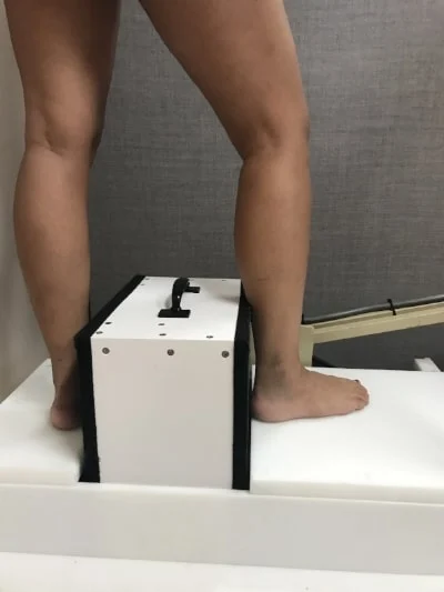
First, a large film cartridge is then be placed next to the limb in question. If the limb is injured, your radiology technicians will work with you to find a comfortable position. If you’re wearing a metal brace of any type you’ll be asked to remove it before any pictures are taken. Even if the area being imaged is far away from the pelvis you’ll still be given a lead apron to shield the groin.
It’s almost certain that multiple images will be taken to show different aspects of the joint or bone. According to many radiologists, “one view is no view.” It’s also likely that images will be taken above or below the site of injury. When taking X-Rays of the knee, for example, pictures of the hip and ankle are often taken as well.
Taking pictures of the unaffected limb for side-to-side comparisons is also very common. This is especially important in children with rapidly developing bones. Only children have growth plates located near the ends of each long bone, e.g. the femur. If these growth plates are fractured, long-term negative effects are likely. Taking side-to-side images is one of the best ways to detect such fractures.
A series of extremity X-Rays typically takes less than 10 minutes. If physical film is being used it takes a further 5 minutes to develop the images. If digital images are being taken they can be viewed on a computer monitor immediately.
Taking X-Rays isn’t painful. At worst, you’ll be mildly uncomfortable lying on a hard X-Ray table for several minutes. If holding your injured limb in position is an issue pillows or sandbags can be used for support.
Potential Risks of Foot X Ray
Almost all medical procedures carry some level of risk. When taking X-Rays, the involved area will be exposed to very low doses of radiation, which can damage living cells. In almost all cases, however, the potential benefits outweigh any risks. It helps to put this risk into perspective. You are exposed to some natural radiation when taking a long plane flight, for example a one way trip from New York to Los Angeles. This small dose is about equal to what you’ll receive during one chest X-Ray.
Interpreting the Results:
X-Ray images are routinely taken of all the major extremities. Commonly visualized areas include the feet, ankles, knees, hips, shoulders, elbows, wrists, and hands. Although it’s an older technology, X-Ray exams remain an extremely good method for evaluating bone and joint injuries. They can also screen for disorders such as arthritis, osteoporosis (bone loss), and bony growths (tumors).
Normal Foot X Ray images:
- No metal or glass fragments or other foreign bodies are noted. The bones, joints, and surrounding soft tissue look unremarkable when compared to “textbook” images.
- Abnormal bony growths, i.e. tumors are not noted. No signs of infection are visible.
- Joints appear properly aligned with no signs of dislocation, arthritis, or other joint conditions.
- Prosthetics such as artificial joints appear normally aligned. Hardware such as rods, plates, and screws appear to be in the correct position.
Abnormal Foot X Ray Images:
- Fractures, including stress or hairline fractures, are present.
- Pieces of metal, glass, or other foreign bodies are noted.
- Abnormal bony growths, i.e. tumors, are noted.
- Common signs of infection are visible. These include collections of fluids such as blood or pus. Gas pockets may also be present.
- A dislocation may be evident.
- Joint or bone damage may be noted from conditions such as arthritis, gout, osteoporosis, Paget’s disease, and many others. Many conditions have tell-tale signs which a radiologist is trained to recognize.
- Swelling may be evident in the periosteum, the soft tissue covering of the bone. This is evident on X-Ray even if the bone itself appears normal.
- Artificial joints or other prostheses are visibly damaged. Hardware such as rods, plates, pins, and screws are not in proper alignment.
Other Considerations:
There are several reasons why you won’t be able to have an X-Ray exam. It’s also possible that you won’t get quality images. These include the following:
- An inability to sit or lay still. Even a small amount of movement can lead to blurry, poor quality images.
- Extreme obesity. Many of the X-Rays are blocked by excess tissue, causing poor quality images. Even if the strength of the beam is turned up, images may still lack detail.
- If you’re unable to get the injured limb into the correct position another imaging technique might be better. A good alternative is Magnetic Resonance Imaging (MRI). Quality X-Ray images depend upon careful positioning of the patient.
- If you are pregnant, or think you might be pregnant, special precautions must be followed. An X-Ray study of the hips or pelvis will have to be rescheduled until after the delivery. Other imaging techniques such as MRI can be used to image pregnant patients.
Foot X Ray Pittsburgh Other Factors:
· If you get a repeat X-Ray exam of the same limb the images may be slightly different. Different medical centers have different equipment and may do things slightly different. If the images are high-quality, however, they’ll still contain the information your doctor needs.
· Foot X-Ray studies aren’t the best at imaging soft tissue structures. These include ligaments, tendons, and cartilage. All of these are key structural components of the body’s joints. A Computed Tomography (CT) or Magnetic Resonance Imaging (MRI) exam are much better at imaging soft tissue.
· Not every extremity injury needs to be imaged. Often the doctor will have a good understanding of what’s going on. Tests like X-Ray exams won’t change the treatment plan.
· It’s possible to miss small fractures or subtle changes on an X-Ray image. Some modalities produce finer detail, such as MRIs, CT scans, and bone scans. If a less finding is suspected, it’s best to use one of the above methods.
Ultrasound of the Foot:
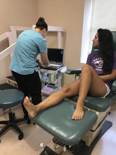
One of the best in office modalities to image the soft tissue of the foot is ultrasound. Tendons, veins, foriegn bodies, hematomas are all well visualized on foot ultrasound exams.
Computed Tomography (CT) Scans:
Computed Tomography (CT) is another imaging technique relies on X-Rays. Unlike traditional X-Rays, it can be used to take pictures of soft tissue structures such as ligaments, tendons, and cartilage. This makes it ideal when joints are being image. It can even image internal organs. CT is one of the most versatile imaging methods available.
The actual process of CT testing somewhat resembles that of Magnetic Resonance Imaging (MRI). The patient lies on a flat table, remains very still, and a donut-shaped device is positioned around the body. X-Ray pulses are generated by this ring, and a thin “slice” image is produced. This scanning ring is adjusted many times to obtain many different images from different angles. CT scans are a good alternative if limb can’t be put into the proper position for an X-Ray.
Digital Foot X Ray
Once all of the images are taken, computer software is used to combine them. The doctor will then scroll through these image to get a more complete view of the joint. This includes all soft tissue structures.
As mentioned, Computed Tomography is also used to visualize organs. These include all major organ systems: the liver, kidneys, heart, lungs, pancreas, spleen, adrenal glands, and colon. When used properly it can even image blood vessels and the spinal cord.
Using a contrast dye makes CT even more versatile. An iodine dye is first injected into the patient. When the CT images are taken highly detailed images of the circulatory system can be obtained. This is extremely useful for assessing the blood flow to an organ or tumor. If the intestines are being imaged then the patient simply drinks the dye.
Magnetic Resonance Imaging (MRI) Exams:
Magnetic Resonance Imaging (MRI) is another advanced imaging technique. Like Computed Tomography (CT) scans, MRI testing is extremely versatile. MRI exams can provide your doctor with different information than CT scans offer. Between MRI and CT scans all structures within the human body can be viewed in very fine detail. Currently MRI exams are the “gold standard” for imaging the body’s joints.
Instead of X-Ray energy, MRI exams use magnetic pulses to obtain images. Much as with CT scans, MRI exams use a highly adjustable donut-shaped ring. In this way many different “slice” images are obtained from many different angles. Also like CT, the patient must lie very still while the MRI exam is taking place.
Just as with CT scans and digital X-Rays, MRI images can be stored on a computer. This means that your doctor has immediate access. The image files can be sent electronically to other doctors for review.
Bone Scans vs Foot X Ray Pittsburgh
Bone scans are specialized tests performed solely to detect bone conditions. In particular, this type of exam helps diagnose and monitor cancer which has spread to the bones. They also give information on processes like infection and arthritis. Unlike other imaging techniques, bone scans show what’s happening inside the bone. They can identify regions which have too much or too little blood flow.
Before a bone scan a tiny amount of radioactive dye is injected into the patient’s veins. It’s not strongly radioactive, and is quickly and thoroughly eliminated from the body. The actual dose of radiation you’ll receive is minimal, just as it is with X-Ray exams. First this “tracer” dye has circulated throughout the body. Then a highly specialized camera is used to identify areas of high (or low) blood flow. Areas where the dye is concentrated show high flow and are called “hot.” Areas with low flow are known as “cold” areas.
Hot areas
This provides your doctor with critical information. “Hot” areas can regions of rapid bone growth, including bone cancer. They also show areas where the bone is being remodeled, as with a healing fracture. “Cold” areas indicate impaired blood flow. This may suggest bone damage such as infection. In all cases, areas of dead or dying (necrotic) bone must be removed as quickly as possible. infection or other damaging processes
Bone scans excel at detecting conditions such as cancer months before other imaging techniques. This allows treatment to begin earlier, and can greatly improve a patient’s outcome.
How long does a typical foot X-ray procedure take?
The duration of a foot X-ray procedure is relatively short. On average, it takes approximately 10 to 15 minutes to complete a foot X-ray. The actual time may vary depending on factors such as the complexity of the imaging required, the patient’s cooperation, and the efficiency of the imaging facility.
During the procedure, the patient will be asked to position their foot as directed by the radiologic technologist, and X-ray images will be taken from different angles to capture a comprehensive view. After the images are obtained, the patient usually receives instructions and can resume normal activities immediately.
Are there any age or health-related considerations for individuals undergoing foot X-rays?
Age and health-related considerations are important factors when undergoing foot X-rays:
1. Pregnancy: It’s crucial for individuals who are pregnant or suspect they might be pregnant to inform their healthcare provider before undergoing any X-ray procedure. While foot X-rays involve a lower radiation dose and may not pose a significant risk, precautions are often taken to minimize exposure during pregnancy.
2. Pediatric Patients: For children, healthcare providers may use different techniques to ensure a minimal radiation dose, considering the increased sensitivity of developing tissues.
3. Underlying Health Conditions: Individuals with certain health conditions, such as circulatory problems or diabetes, may need special considerations during foot X-rays to ensure their safety and well-being.
Can X-rays distinguish between different types of tissues in the foot, such as bones and soft tissues?
X-rays are primarily used to visualize bones and are not as effective at distinguishing between different types of soft tissues in the foot. In X-ray images, bones appear as dense structures and are easily visible due to their ability to absorb X-rays. However, soft tissues, such as muscles, tendons, and ligaments, are not as dense and may not be well-differentiated in standard X-ray images.


