
Digital Mucoid Cyst

What is a Digital Mucous Cysts (DMC)?
Overview, signs, and symptoms
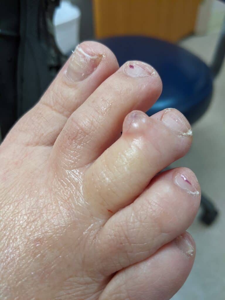
Digital mucous cysts are benign, fluid-filled cysts commonly found on the fingers. Specifically, they are most often located at the proximal nail fold of the distal interphalangeal joints. In plain English, this means they usually occur on the skin that borders the bottom of the fingernails. Among medical practitioners simply refer to them by their abbreviation, DMCs. By definition, digital mucous cysts occur on the hands, yet similar cysts may also appear on the toes.
In most cases, Digital mucous cysts are asymptomatic, if unsightly. This means that they don’t actually bother the patient beyond cosmetic concerns. Various minimally-invasive procedures are employed when treatment is required. Even with treatment, the recurrence of problematic cysts is common.

Medical science is unsure as to the exact cause of DMCs. Yet while this is true, mucoid degeneration of the affected tissue is suspected (more on this later). Unfortunately, research into this common condition has been far from extensive. This is primarily due to the asymptomatic nature of most DMCs. To complicate matters, digital mucous cysts are referred to by many different names in medical literature. These include:
- Cystomatas
- Periarticular fibromas
- Periungual ganglions
- Myxoid or mucus cysts
- Dorsal nail cysts
- Myxomatous degenerative cysts
- Digital mucinous pseudocysts
While all of these terms refer to the same condition, “digital mucous cysts” is the most widely used.
Etiology (cause) of Digital Mucoid Cyst
As mentioned, the precise mechanism behind the formation of DMCs remains elusive. Connective tissue degeneration of the proximal nail fold is currently thought to be responsible. Direct physical trauma to the nail or joint may also be a contributing factor. In most cases, the cyst connects with the synovial space of the distal interphalangeal joint (DIP). This makes them less of a surface phenomenon, and may contribute to arthritis and spurring of the joint. When DMCs do cause the patient discomfort this is usually what’s at play.
Prognosis
DMCs don’t typically pose any real threat to patients, hence have a good prognosis. They may return, however, unless invasive surgery is undertaken. While such radical procedures are effective they’re also riskier, so less-invasive options are attepted first. In some cases the cyst may impinge upon adjacent nerve fibers, causing pain. Deformation of the affected nail can also occur if the cyst is large enough.
Medical History of Digital Mucous Cysts (DMCs)
As mentioned previously, most DMCs cause no symptoms whatsoever. The time it takes for them to develop varies considerably between patients. Some cysts appear suddenly, while others manifest slowly over many months. Often the affected nail will take on a grooved appearance before the cyst itself appears. Osteoarthritis, also known as “wear and tear” arthritis, frequently occurs in the joint closest to the cyst. The majority of cysts remain until removed, yet a significant minority disappear without intervention.
The reason why cysts appear in some patients and not others remains unclear. Currently there are no known risk factors which predict DMCs. Another way of putting this is that there are no “typical” DMC patients. Previous trauma to the affected digit, however, is noted in a small percentage of cases. Patients are more likely to seek medical attention for larger cysts, which may cause discomfort or outright pain. Also note that large DMCs can affect the patient’s ability to use the affected finger properly.
A typical digital mucous cyst is round to oval in shape with a raised, domed profile. They range anywhere from tiny (1 mm) to very large (>10 mm). The cysts in the second, larger size bracket are almost exclusively responsible for bothersome symptoms. This primarily consists of pain and altered function of the finger. Technically, any finger may develop a DMC. In clinical practice, cysts are most often seen on the index or middle finger of the patient’s dominant hand. Digital mucous cysts contain a thick, gelatinous fluid which may be clear or slightly yellowish.
Are Digital Mucoid Cyst harmful?
While they are generally not harmful, they can cause discomfort and may affect daily activities depending on their size and location.
Digital mucoid cysts usually appear as small, fluid-filled bumps near the nail bed, and they may be associated with osteoarthritis or degenerative joint disease. The exact cause of these cysts is not fully understood, but they are believed to develop from disruptions in the joint capsule or adjacent connective tissue.
In most cases, digital mucoid cysts do not require immediate medical intervention. However, if they cause pain, interfere with hand function, or become infected, medical attention may be necessary. If the cyst becomes infected, it can lead to redness, swelling, tenderness, and pus drainage. In such cases, a healthcare professional may recommend treatment options such as antibiotics or drainage.
If a digital mucoid cyst is causing significant discomfort or affecting your quality of life, a healthcare professional may suggest intervention through procedures like needle aspiration, where the fluid is drained from the cyst, or surgical removal. These procedures aim to alleviate symptoms and prevent recurrence.
It’s important to consult with a healthcare professional, such as a dermatologist or hand specialist, to properly evaluate and determine the best course of action for your specific situation. They can provide a thorough assessment, offer treatment options, and address any concerns you may have regarding the digital mucoid cyst.
What to do if you gain one?
If you notice a digital mucoid cyst and it is not causing significant discomfort or interfering with your daily activities, you may choose to monitor it for changes. However, if the cyst becomes painful, infected, or bothersome, it is advisable to seek medical attention.
A healthcare professional, such as a dermatologist or hand specialist, can evaluate the cyst and recommend appropriate treatment options, which may include draining the fluid through needle aspiration or surgical removal.