
Broken Ankle

Broken Ankle
Doctor Examination of a broken ankle
Prior to treating your broken ankle, your doctor will discuss your medical history with you. This will include a review of symptoms and mechanism of injury. A physical examination of the ankle, foot, and lower leg will follow.
Following discussion of your medical history including symptoms and how the injury was caused, your doctor will do an examination of your ankle, foot, and lower leg.
Imaging Tests
An X-ray examination is usually needed to rule in or rule out a fracture. By far, this is the most commonly used imaging modality used in this fashion. It allows your doctor to clearly see hard tissue (bone), and can identify any visible misalignments. These are known as dislocations.
Yet however useful X-ray is it suffers from a major drawback. Soft tissue structures such as ligaments and tendons aren’t visible on X-Ray imaging. Other tests must be used, such as Magnetic Resonance Imaging (MRI) and CT (Computed Tomography) Scans.
XRAYS
If your doctor suspects that your ankle is fractured, he or she will make the request for other tests to be performed to gain further information. X-rays rank as the most commonly used form of diagnostic imaging. It allows the radiologist to identify if the bone has been broken or displaced. Also, an x-ray can identify how many pieces of the bone are broken. X-rays may be taken of the leg, ankle, and foot to ensure that other areas are not injured.
Physical Exam
Note that parts of the physical examination can be used in conjunction with X-Ray. This is known as a “stress test.” The goal of this type of exam is to determine if an ankle fracture requires surgical intervention.
Under different circumstances, your healthcare provider may apply pressure on the ankle joint and take a different x-ray, called a stress test. This form of x-ray is performed to identify whether the ankle fractures require surgery or not.
CT
If there’s any doubt, a CT Scan is usually the next step. This imaging technique has several advantages. Firstly, it produces high-quality cross-sectional “slice” images, much like an MRI study. Secondly, CT Scans visualize soft tissue structures quite well. This makes it valuable for severe sprains which extend deeply into the ankle joint when a fracture is suspected. Thirdly, this modality costs less than an MRI.
MRI
MRI, known as Magnetic Resonance Imaging, provides high resolution images of bones, as well as soft tissues such as ligaments. It may also be done for the sole purpose of evaluating the ankle ligaments.
Fractures can occur at different levels of the fibula. This will influence the route of treatment.
Nonsurgical Treatment
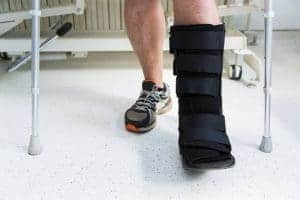
Methods for protection of the ankle include wearing high-top tennis shoes or a short leg cast. A physician may allow weight on the leg right away, but others may choose to wait for six weeks.
Physicians will see you regularly to reexamine your ankle on the x-ray. This allows them to see if fragments of the fracture have moved out of place.
Surgical Treatment
Stable ankles may not require surgery. The bone could be broken but not out of place or barely out of place. If the ankle is stable there may not be a need for a stress x-ray. Treatment can vary based on the location of the break.
Surgery is needed if an ankle is out of place and unstable. Bones of the ankle are repositioned to normal alignment at the start of surgery. Metal plates and special screws are attached to the outer portion of the bones to hold them together. Similarly, screws or rods may be used to hold bone fragments in place until healed.
Treatment: Medial Malleolus Fracture
A fracture of the medial malleolus is a fracture on the inside portion of the lower leg. These can occur at different levels as well. These fractures can occur in conjunction with a lateral malleolus, posterior malleolus, or with ligament injuries of the ankle.
Nonsurgical Treatment
Fractures that are not out of place or are very minor may be treated without a surgical procedure. This may be ruled out on a stress x-ray to determine the outcome. The fracture may be treated with a short leg cast or a removable brace. Usually, you need to avoid putting weight on your leg for approximately 6 weeks. Repeated x-rays will be performed to view the progress of treatment. Later you will do physical therapy.
Surgical Treatment of a broken ankle
Surgery may be recommended if the ankle is out of place/unstable.
The left X-ray is of the medial malleolus fracture. X-ray on the right is a surgical repair with a plate and screws.
Surgery may be considered although the fracture is not out of place. This is done to reduce the risk of fractures not healing and to allow the ankle to move earlier.
Fractures of the medial malleolus may include impaction or indentation of the ankle joint. When a force is so great that it drives one end of a bone into another it is called impaction. This may require bone grafting to repair impacted fractures. Bone fragments can also be repaired with screws, plates, and wiring techniques.
Treatment: Posterior Malleolus Fracture
This fracture occurs at the back of the tibia at the lower portion at the ankle joint.
It often occurs that the lateral malleolus (fibula) is broken along with the posterior malleolus. This is because they share common ligament attachments. A medial malleolus fracture can also occur.
If the broken piece on the posterior malleolus is big, the ankle may be unstable. Studies support that the bigger the piece is, the more unstable the ankle is and should seek treatment via surgery.
To prevent the risk of arthritis, the podiatrist should diagnose and treat a posterior malleolus fracture . The posterior tibia contains a lot of cartilage where the bone breaks. This smooth surface lines the joint and if the broken bone is larger than 25% of the ankle as well as being out of place more than a few millimeters, the cartilage will not heal properly. This uneven surface may lead to uneven pressure on the joint surface, which can further lead to arthritis.
Nonsurgical Treatment
Fractures have the ability to be treated without surgery.
Treatment may involve a short leg cast or removable brace. Patients are typically told not to bear weight on the ankle for at least 6 weeks.
Surgical Treatment
The ankle may be out of place and require surgery.
Options of screw placement could be placed from the front of the ankle to the back, or vise versa. Lastly, a plate and screws may also be placed along the back of the tibia.
Treatment: Bimalleolar Fractures or Bimalleolar Equivalent Fractures
Bimalleolar refers to the two of three parts of the malleoli being broken.
Most often, the lateral and medial malleolus are broken which result in an unstable ankle.
A bimalleolar equivalent fracture means that the medial ligaments are injured along with the malleoli being broken.
This can be identified on a stress test x-ray.
Both types of fractures are unstable for the patient and can be associated with a dislocation.
Nonsurgical Treatment
These injuries are usually unstable and require surgery.
Nonsurgical treatment may be considered if a patient has significant health problems.
Splints are typically provided to immobilize the ankle as the swelling decreases. A short leg cast is usually the next step and may be changed frequently as the swelling subsides.
Follow up with your physician regularly to repeat and interpret x-rays.
Are There Risks Of Re-injury After The Broken Ankle Has Healed?
Yes, after the broken ankle heals there will be an increased risk of Re-injury, especially if it hasn’t fully healed or regained it’s strength, flexibility or alignment. Furthermore, there are more factors like old joint damage, weakened muscles in the surrounding area, and returning to high-impact activities too early can also increase the risks. Therefore, it’s important to get physical therapy to help rebuild strength and stability, this will highly lower the chances of re-injury. You will need to make sure you have a proper footwear for a good recovery too, this will. Also highly reduce the risks of injury.
Will Broken Ankles Interfere With My Mobility For Long-term?
Yes, unfortunately broken ankle can impact on your mobility for a long time, especially if the injury was too serious, too deep or if the healing wasn’t optimal. You can expect problems like stiffness, chronic pain, reduced range of motion, or even arthritis in few cases. Luckily, with proper treatments, rehabilitation, and physical therapy, you should be able to regain full or almost old mobility back! Which is why it’s important to follow the treatment plan and follow your doctor’s post treatment tips.
When Am I Allowed To Start Putting Weight On My Broken Ankle?
When you should be allowed to put weight on your broken ankle depends on the severity of the fracture and the treatment used. Most of the time, you won’t be allowed to engage in eight-bearing activities for 6-8 weeks to allow proper healing, especially if the cast or surgery was involved in it. Your doctor will recommend you to use crutches before you transition to full weight. You must make sure you rest as much as possible and do not try anything that harms your ankle further, otherwise the healing process can become much complicated.
What exercises can help with ankle recovery?
Exercises play a crucial role in ankle recovery after an injury or a broken ankle. They help improve strength, stability, and flexibility of the ankle joint. However, it’s essential to consult with a healthcare professional or a physical therapist before starting any exercise program to ensure they are appropriate for your specific condition. Here are some common exercises that can aid in ankle recovery:
- Ankle Circles:
Sit or lie down with your leg extended. Slowly move your ankle in circular motions, both clockwise and counterclockwise. This helps improve ankle flexibility.
- Alphabet Writing:
Similar to ankle circles, imagine you are writing the alphabet with your toes. This exercise promotes ankle mobility.
- Ankle Pumps:
Sit with your legs extended. Move your ankle up and down, flexing and pointing your toes. This helps with ankle flexibility and blood circulation.
- Resistance Band Exercises:
Use a resistance band tied to a stable object and looped around your forefoot. Move your foot inward, outward, up, and down against the resistance. This improves ankle strength and stability.
- Calf Raises:
Stand with feet shoulder-width apart and rise up on your toes, then lower back down. This exercise strengthens the calf muscles and helps with ankle stability.
- Toe Tapping:
Sit with your feet flat on the floor. Tap your toes up and down quickly for a set period. This helps with ankle strength and flexibility.
- Balance Exercises:
Stand on one leg with the support of a stable object if needed. Try to maintain balance for as long as possible. This improves ankle stability and proprioception.
- Heel Slides:
Sit with your legs extended, and slide your heel along the floor towards your body, bending the knee. Then slide it back to the starting position. This exercise enhances ankle mobility.
Ankle Fractures (Broken Ankle)
A broken ankle is also known as an ankle “fracture.” To meet this criteria, one or more of the bones that make up the ankle joint must be broken. A fracture can be as mild as a simple break in one bone which may not stop you from walking. Unfortunately, a fracture can also mean several fractures which can force the ankle out of place and may require non-weight bearing recovery for a few months. As you may assume, the more bones that are broken will cause the ankle to be increasingly unstable. Also, the ligaments may be damaged which are responsible for holding the bones and ankle joint in the correct position.
Broken ankles can affect people of all ages and activities. Due to an increase in activity of the older population, doctors have noted an increase in the number of ankle fractures over the past 30 to 40 years.
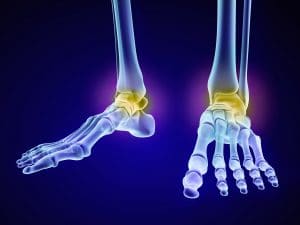
Anatomy
Three bones make up the ankle joint:
- Tibia – shinbone
- Fibula – smaller bone of the lower leg
- Talus – a small bone that sits between the heel bone (calcaneus) and the tibia and fibula
The tibia and fibula have specific parts that make up the ankle:
- Medial malleolus – inside part of the tibia
- Posterior malleolus – back part of the tibia
- Lateral malleolus – end of the fibula
Doctors will classify ankle fractures according to the area of the bone that is broken. If one fractured the end of the fibula, it would be diagnosed as a lateral malleolus fracture. Similarly, if both the tibia and fibula are broken, it will be diagnosed as a bimalleolar fracture.
Two joints are involved in ankle fractures:
- Ankle joint – where the tibia, fibula, and talus meet
- Syndesmosis joint – the joint between the tibia and fibula, which is held together by ligaments
Multiple ligaments help make the ankle joint stable.
Causes of a broken ankle
- Twisting or rotating your ankle
- Rolling your ankle
- Tripping or falling
- Impact during a car accident
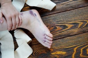
Symptoms
Because a severe ankle sprain can feel the same as a broken ankle, every ankle injury should be evaluated by a physician.
Common symptoms for a broken ankle include:
- Immediate and severe pain
- Swelling
- Bruising
- Tender to touch
- Cannot put any weight on the injured foot
- Deformity (“out of place”), particularly if the ankle joint is dislocated as well